Potency Ranking of Dermal Sensitizing Chemicals Using the IVSA and epiCS® Skin Tissues.
In validation studies at MB Research Labs, we have demonstrated IL-18 release in 3D tissue constructs of reconstructed human epidermis (RHE) in response to dermal sensitizers.
The exposure concentrations resulting in a greater than a threshold positive response (SI ≥ 2.0) correlates with their potency in the In Vitro Sensitization Assay (IVSA). In our experiments, NBB and DNCB were strong inducers of IL-18 secretion (EC2.0 = 0.028% and 0.03%). Isoeugenol (IE) and Cinnamaldehyde (CA) were moderate sensitizers, while Resorcinol and HCA (EC2.0 = 22%) were weak sensitizers. Sensitizer potency ranked as follows: NBB > DNCB > PPD, IE ≈ CA > RES > HCA, with NBB, DNCB and PPD classified as strong, IE and CA as moderate, and RES and HCA classified as weak sensitizers.
Of the 20 chemicals tested, 7 were irritants, 2 were non-sensitizers and only Chlorobenzene was incorrectly predicted as a weak sensitizer.
In summary, measuring IL-18 release from RHE allows for highly accurate and sensitive identification of dermal sensitizers.
INTRODUCTION
The CDC estimates more than 13 million individuals in the US are likely exposed to chemicals that cause skin diseases. Many of these skin diseases are caused by contact dermatitis, a common type of illness associated with many occupational hazards, costing an estimated one billion dollars annually in healthcare and lost productivity (http://www.cdc.gov/niosh/topics/skin/). Currently there are no fully validated and r
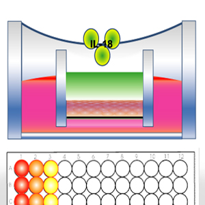
egulatory accepted animal-alternative methods available to assess for Allergic Contact Dermatitis (ACD) / sensitization. In ACD, keratinocytes in the skin are the first to contact and elicit a response to allergens during an exposure. The 3D human epidermis equivalent epiCS® is reconstructed from normal human primary epidermal keratinocytes. Keratinocytes have been shown to secrete a wide range of cytokines. Evidence demonstrates that cytokine IL-18 is an essential component of dermal sensitization. Most notable it has been shown using IL-18-deficient mice that IL-18 is not required for irritation contact dermatitis, but is required for an optimal ACD response (Antonopoulos et al., 2008. Journal of Leukocyte Biology 83: 361-7).
An IL-18 endpoint has been used to predict sensitization in tissue models using filter paper applications (Gibbs et al., 2013, Toxicology and Applied Pharmacology 272(2):529-41). To identify sensitizing compounds, we measured IL-18 secretion from epiCS® after treatment with pure chemical s via direct chemical application.
CONCLUSIONS
An SI of 2.0 was calculated to be the best fit cut-off to discriminate sensitizers from irritants and non-toxic chemicals. The epiCS® IVSA correctly predicted sensitization potential with 89% Accuracy and 89% Sensitivity (18 chemicals).
TV50 and IL-18 SI-2 data obtained from epiCS® tissues had very a high correlation
(r<sup>2</sup>=0.94).
Using an SI-2 in this IVSA, data correlated better with Human DSA05
data (r<sup>2</sup>=0.96) than with LLNA EC3 data (r2=0.81).
For more info about this study and it’s ability to aid you in your sensitization screening, please see: http://www.mbresearch.com/ivsa.htm